Contents
Overview
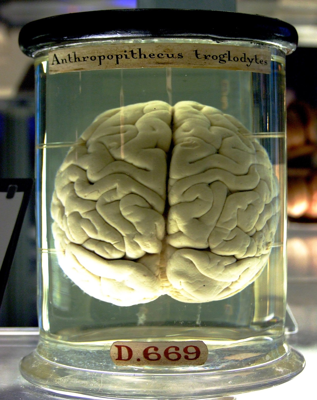
Brain structure refers to the physical organization and arrangement of the nervous tissue that constitutes the brain, the central organ of the nervous system. This intricate architecture dictates how information is processed, stored, and transmitted, underpinning everything from basic reflexes to complex cognition. Understanding brain structure is fundamental to neuroscience, providing the physical basis for behavior, thought, and emotion. It encompasses macroscopic features like distinct lobes and nuclei, as well as microscopic arrangements of nerve cells and glia.
🧭 Key Brain Regions & Their Functions
The brain is broadly organized into several key regions, each with specialized functions. The cerebrum, the largest part, is divided into two hemispheres and four lobes (frontal, parietal, temporal, occipital), responsible for higher-level functions like language, memory, and reasoning. The cerebellum, located at the back, coordinates movement and balance. The brainstem connects the cerebrum and cerebellum to the spinal cord, controlling vital autonomic functions like breathing and heart rate. Deeper structures like the thalamus and hypothalamus play crucial roles in relaying sensory information and regulating bodily homeostasis.
🔬 Microscopic Anatomy: Neurons & Glia
At the microscopic level, the brain is composed of billions of nerve cells, the primary signaling units, and a diverse population of glia, which provide support, insulation, and nourishment. Neurons communicate via electrical and chemical signals across specialized junctions called synapses. Glial cells, including astrocytes, oligodendrocytes, and microglia, are essential for maintaining the neuronal microenvironment, facilitating synaptic transmission, and protecting the brain from injury and disease. This intricate cellular network forms the basis of all neural computation.
📈 Development & Plasticity
Brain structure is not static; it undergoes significant development from prenatal stages through adulthood and exhibits remarkable neuroplasticity. Early development involves neurogenesis (the birth of new neurons) and neuronal migration to form distinct brain regions. Throughout life, experiences can modify neural connections, strengthening or weakening synapses, and even generating new neurons in specific areas like the hippocampus. This plasticity allows the brain to adapt to changing environments and learn new information.
💡 Research & Tools
Investigating brain structure relies on a sophisticated toolkit. Neuroimaging techniques like Magnetic Resonance Imaging (MRI) and Positron Emission Tomography (PET) allow non-invasive visualization of brain anatomy and function. Electrophysiology measures electrical activity, while histology and electron microscopy enable detailed examination of cellular and subcellular structures. Computational neuroscience uses mathematical models to simulate neural networks and understand how their structure gives rise to function.
🤔 Debates in Brain Structure
Significant debates persist regarding brain structure. One major area of contention is the extent to which structure is genetically determined versus shaped by experience, particularly during critical developmental periods. Another is the precise relationship between specific neural circuits and complex cognitive functions like consciousness or decision-making. The role of microglia in neurodegenerative diseases, once thought purely supportive, is now a hotbed of research, with evidence suggesting active participation in disease processes.
🌟 Clinical Significance
Understanding brain structure is critical for diagnosing and treating a wide range of neurological and psychiatric disorders. Structural abnormalities can underlie conditions such as stroke, traumatic brain injury, Alzheimer's disease, and schizophrenia. By mapping structural changes, clinicians can better understand disease progression, predict patient outcomes, and develop targeted therapeutic interventions, including deep brain stimulation and pharmacological treatments.
🚀 Future Directions
The future of brain structure research promises even deeper insights. Advances in connectomics aim to map the complete wiring diagram of the brain, revealing the intricate network of connections between neurons. Optogenetics and other cutting-edge tools allow unprecedented control over neural activity, enabling researchers to probe causal relationships between structure and function. Ultimately, this work could lead to novel strategies for repairing damaged brains and enhancing cognitive capabilities.
Key Facts
- Year
- 2023
- Origin
- Scientific Research
- Category
- Neuroscience
- Type
- Scientific Concept
Frequently Asked Questions
What are the main parts of the brain?
The brain is typically divided into the cerebrum (responsible for higher functions), the cerebellum (coordinating movement), and the brainstem (controlling vital functions). Each of these major divisions is further subdivided into lobes and nuclei with specialized roles. Deeper structures like the thalamus and hypothalamus also play critical roles in sensory processing and regulation.
How do neurons communicate?
Neurons communicate through electrochemical signals. An electrical impulse travels down the neuron's axon, triggering the release of chemical messengers called neurotransmitters at the synapse. These neurotransmitters then bind to receptors on the next neuron, potentially initiating a new electrical signal.
Can brain structure change after childhood?
Yes, absolutely. The brain exhibits significant neuroplasticity throughout life. While major structural development occurs early, experiences, learning, and even injury can lead to changes in synaptic strength, the formation of new connections, and in some areas, the birth of new neurons.
What is the difference between gray matter and white matter?
Gray matter consists primarily of neuronal cell bodies, dendrites, and unmyelinated axons, and is where information processing occurs. White matter is mainly composed of myelinated axons, which act as the brain's 'wiring,' transmitting signals rapidly between different gray matter regions. Myelin, a fatty substance, gives white matter its characteristic color.
How do scientists study brain structure?
Scientists use a variety of methods, including neuroimaging techniques like MRI and PET scans to view the living brain, histology to examine tissue under a microscope, and electrophysiology to record neural activity. Computational models are also used to simulate and understand neural networks.
What is the role of glial cells?
Glial cells are crucial support cells in the brain. They provide structural support, insulate neurons with myelin, supply nutrients, remove waste products, and play active roles in synaptic function and immune responses within the brain. Different types of glia, like astrocytes and microglia, have distinct functions.